


R&D: Scientists Catch Light Squeezing and Stretching Next-Gen Storage Material
Combining X-ray and electron data from two cutting-edge SLAC instruments, researchers observe rapid atomic response of iron-platinum nanoparticles to light, which could help control future magnetic storage devices.
This is a Press Release edited by StorageNewsletter.com on January 30, 2018 at 2:21 pmBy Manuel Gnida, SLAC National Accelerator Laboratory, Department of Energy
Scientists at the Department of Energy’s SLAC National Accelerator Laboratory have seen for the first time how atoms in iron-platinum nanoparticles – a next-generation material for magnetic data storage devices – respond extremely rapidly to brief laser flashes.
New information on the ultrafast atomic response of iron-platinum nanoparticles
to laser light could help researchers develop ways to manipulate and control
next-generation magnetic data storage devices with light.
The data were obtained with a unique combination of SLAC’s Linac
Coherent Light Source (LCLS) X-ray laser and instrument
for ultrafast electron diffraction (UED).
(Greg Stewart/SLAC National Accelerator Laboratory)
Understanding these fundamental motions could potentially lead to new ways of manipulating and controlling such devices with light.
Combining snapshots from two world-leading ultrafast atomic-resolution ‘cameras’ at SLAC – the Linac Coherent Light Source (LCLS) X-ray laser and an apparatus for ultrafast electron diffraction (UED) – the team showed that the laser flashes demagnetized the iron-platinum particles within less than a trillionth of a second, causing atoms in the material to move closer together in one direction and move further apart in another.
The results also provide the first atomic-level description of the mechanical strain, known as magnetostriction, occurring in magnetic materials when the magnetization is changed. The phenomenon manifests itself in many ways, including the electric hum of transformers. Before the study, published in Nature Communications, researchers had assumed that these structural changes happen relatively slowly. However, the new data suggest that ultrafast processes could play an important role.
“Previous models of the properties of iron-platinum nanoparticles did not consider these extremely fast and fundamental atomic motions,” says Hermann Dürr, study’s principal investigator, Stanford Institute for Materials and Energy Sciences (SIMES), which is jointly operated by SLAC and Stanford. “Although we don’t yet understand the full ramifications of these processes, including them in our calculations could open up new pathways for the development of future data storage technologies.”
Pushing limits of magnetic data storage
Magnetic storage devices are widely used to record information produced in virtually all areas of our digital world, and they are believed to remain crucial data storage solutions for the foreseeable future. Faced with ever growing amounts of global data volumes, hardware engineers are aiming to maximize the density with which these media can store information.
However, current technologies are coming close to their technical limits. Today’s hard disk drives, for example, can reach storage densities of several hundred billion bits per square inch, and similar future devices aren’t expected to exceed much more than a trillion bits per square inch. New developments are required to take magnetic data storage to the next level.
“A very promising approach that could take us there is heat-assisted magnetic recording in hard drives using nanosized grains of materials like iron-platinum,” says Eric Fullerton, director, Center for Memory and Recording Research, University of California, San Diego, CA, and a co-author of this study. “In this method, the information is encoded with a nanofocused laser and a magnetic field, or possibly even a laser alone, that switch the magnetization of the nanoparticles. These next-generation drives, which can have much larger storage densities, are already being tested in industry and could soon become commercially available.”
The SLAC study looked into an important facet of this technology – the interaction of laser light with iron-platinum nanoparticles.
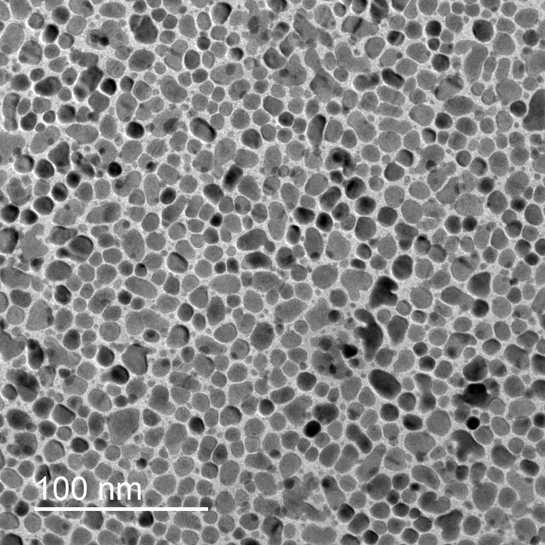
Image of iron-platinum nanoparticles taken
with a scanning transmission electron microscope
(Tyler Chase/Stanford/SLAC National Accelerator Laboratory)
Combining X-Ray and electron vision
The researchers first took the nanoparticles, which were about 50 atoms in diameter, to LCLS, where they shone a brief optical laser pulse onto them. With LCLS’s ultrabright femtosecond X-ray flashes they were able to follow how the laser light changed the magnetization of the material – from completely magnetized to largely demagnetized. One femtosecond is a millionth of a billionth of a second.
They repeated the experiment using the UED instrument at SLAC’s Accelerator Structure Test Area (ASTA), in which a pulsed beam of very energetic electrons probed the samples. With this method, the scientists made a stop-motion movie of how atoms in the nanoparticles moved after being hit by laser light.
“Only the combination of both methods allowed us to see the full picture of the ultrafast atomic response to laser light,” says Alexander Reid, study’s lead author, SIMES and LCLS. “The laser pulse alters the magnetization in the material, which, in turn, drives structural changes and causes mechanical strain.”
Xijie Wang, head, SLAC’s UED initiative, says, “This study demonstrates how powerful the two methods are together. The high-energy electron beam was absolutely crucial in the determination of the 3-D atomic motions, and without X-rays we wouldn’t have been able to link these motions to the material’s magnetic behavior.”
In addition to SLAC and Stanford researchers, the collaboration included scientists from the Czech Republic, Germany, Japan, Sweden, The Netherlands, as well as several U.S. institutions. Part of this study was supported by the DOE Office of Science and the Laboratory Directed Research and Development (LDRD) Program. LCLS is a DOE Office of Science user facility.
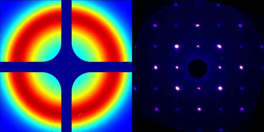
Intensity pattern on a detector created by X-rays (left) and electrons
that have passed through a sample of iron-platinum nanoparticles.
The X-ray data reveal information about the sample’s magnetic state,
and the electron data provide details of the atomic structure.
(Alexander Reid/SLAC National Accelerator Laboratory)
SLAC is a multi-program laboratory exploring frontier questions in photon science, astrophysics, particle physics and accelerator research. Located in Menlo Park, CA, SLAC is operated by Stanford University for the U.S. Department of Energy’s Office of Science.
SLAC National Accelerator Laboratory is supported by the Office of Science of the U.S. Department of Energy. The Office of Science is the single largest supporter of basic research in the physical sciences in the United States, and is working to address some of the most pressing challenges of our time.
Article : Beyond a phenomenological description of magnetostriction
Nature Communications has published an article written by A. H. Reid, Stanford Institute for Materials and Energy Sciences, SLAC National Accelerator Laboratory, 2575 Sand Hill Road, Menlo Park, CA, 94025, USA, and Linac Coherent Light Source, SLAC National Accelerator Laboratory, 2575 Sand Hill Road, Menlo Park, CA, 94025, USA, X. Shen, Accelerator Division, SLAC National Accelerator Laboratory, 2575 Sand Hill Road, Menlo Park, CA, 94025, USA, P. Maldonado, Department of Physics and Astronomy, Uppsala University, P. O. Box 516, S-75120, Uppsala, Sweden, T. Chase, Stanford Institute for Materials and Energy Sciences, SLAC National Accelerator Laboratory, 2575 Sand Hill Road, Menlo Park, CA, 94025, USA, and Department of Applied Physics, Stanford University, Stanford, CA, 94305, USA, E. Jal, Stanford Institute for Materials and Energy Sciences, SLAC National Accelerator Laboratory, 2575 Sand Hill Road, Menlo Park, CA, 94025, USA, and CNRS, Laboratoire de Chimie Physique – Matière et Rayonnement, Sorbonne Universités, UPMC Univ. Paris 06, 75005, Paris, France, P. W. Granitzka, Stanford Institute for Materials and Energy Sciences, SLAC National Accelerator Laboratory, 2575 Sand Hill Road, Menlo Park, CA, 94025, USA, Van der Waals-Zeeman Institute, University of Amsterdam, 1018XE, Amsterdam, The Netherlands, K. Carva, Faculty of Mathematics and Physics, Department of Condensed Matter Physics, Charles University, Ke Karlovu 5, CZ-12116, Prague 2, Czech Republic, R. K. Li, Accelerator Division, SLAC National Accelerator Laboratory, 2575 Sand Hill Road, Menlo Park, CA, 94025, USA, J. Li, L. Wu, Brookhaven National Laboratory, Upton, NY, 1193, USA, T. Vecchione, Accelerator Division, SLAC National Accelerator Laboratory, 2575 Sand Hill Road, Menlo Park, CA, 94025, USA, T. Liu, Stanford Institute for Materials and Energy Sciences, SLAC National Accelerator Laboratory, 2575 Sand Hill Road, Menlo Park, CA, 94025, USA, and Department of Physics, Stanford University, Stanford, CA, 94305, USA, Z. Chen, Stanford Institute for Materials and Energy Sciences, SLAC National Accelerator Laboratory, 2575 Sand Hill Road, Menlo Park, CA, 94025, USA, and Department of Physics, Stanford University, Stanford, CA, 94305, USA, D. J. Higley, Stanford Institute for Materials and Energy Sciences, SLAC National Accelerator Laboratory, 2575 Sand Hill Road, Menlo Park, CA, 94025, USA, and Department of Applied Physics, Stanford University, Stanford, CA, 94305, USA, N. Hartmann, R. Coffee, Linac Coherent Light Source, SLAC National Accelerator Laboratory, 2575 Sand Hill Road, Menlo Park, CA, 94025, USA , J. Wu, Accelerator Division, SLAC National Accelerator Laboratory, 2575 Sand Hill Road, Menlo Park, CA, 94025, USA, G. L. Dakovski, W. F. Schlotter, Linac Coherent Light Source, SLAC National Accelerator Laboratory, 2575 Sand Hill Road, Menlo Park, CA, 94025, USA, H. Ohldag, Stanford Synchrotron Radiation Laboratory, SLAC National Accelerator Laboratory, 2575 Sand Hill Road, Menlo Park, CA, 94025, USA, Y. K. Takahashi, Magnetic Materials Unit, National Institute for Materials Science, Tsukuba, 305-0047, Japan, V. Mehta, San Jose Research Center, HGST a Western Digital Company, 3403 Yerba Buena Road, San Jose, CA, 95135, USA, and Thomas J. Watson Research Center, 1101 Kitchawan Road, Yorktown Heights, NY, 10598, USA, O. Hellwig, San Jose Research Center, HGST a Western Digital Company, 3403 Yerba Buena Road, San Jose, CA, 95135, USA, Institute of Physics, Technische Universität Chemnitz, Reichenhainer Straße 70, D-09107, Chemnitz, Germany, and Institute of Ion Beam Physics and Materials Research, Helmholtz-Zentrum Dresden–Rossendorf, 01328, Dresden, Germany, A. Fry, Linac Coherent Light Source, SLAC National Accelerator Laboratory, 2575 Sand Hill Road, Menlo Park, CA, 94025, USA, Y. Zhu, Brookhaven National Laboratory, Upton, NY, 1193, USA, J. Cao, Department of Physics and National High Magnetic Field Laboratory, Florida State University, Tallahassee, FL, 32310, USA, E. E. Fullerton, Center for Memory and Recording Research, UC San Diego, 9500 Gilman Drive, La Jolla, CA, 92093-0401, USA, J. Stöhr, Stanford Institute for Materials and Energy Sciences, SLAC National Accelerator Laboratory, 2575 Sand Hill Road, Menlo Park, CA, 94025, USA, P. M. Oppeneer, Department of Physics and Astronomy, Uppsala University, P. O. Box 516, S-75120, Uppsala, Sweden, X. J. Wang, Accelerator Division, SLAC National Accelerator Laboratory, 2575 Sand Hill Road, Menlo Park, CA, 94025, USA, and H. A. Dürr, Stanford Institute for Materials and Energy Sciences, SLAC National Accelerator Laboratory, 2575 Sand Hill Road, Menlo Park, CA, 94025, USA, and Department of Physics and Astronomy, Uppsala University, P. O. Box 516, S-75120, Uppsala, Sweden.
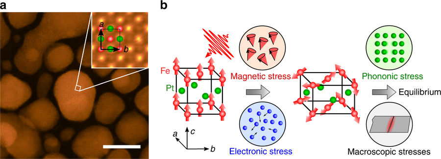
FePt physical structure and the time sequences of contributions to the lattice strain.
a High-angle annular dark-field scanning transmission electron microscopy images
of the morphology and a-, b-plane atomic arrangement (inset) for FePt nanoparticles
embedded in an amorphous carbon matrix. 10 nm scale bar shown in lower right.
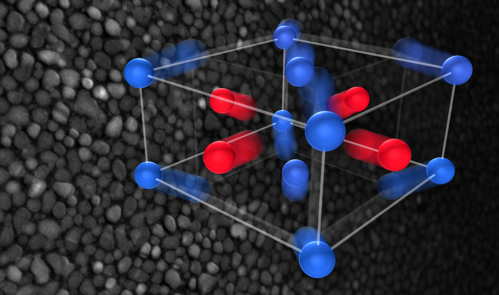
b The FePt crystal structure and magnetic arrangement. Fe and Pt atomic positions
in the L10 lattice structure are indicated by red and green spheres
in the insets, respectively. The majority of the grains are aligned with the a and b unit
cells directions in the plane of the film. The schematic shows the sequence of stress
contributions in FePt. In the first picosecond after laser excitation, the lattice
is subject to the rapidly developing magnetic and electric stresses.
This leads to an increase in the tetragonal distortion of the unit cell as illustrated.
On a timescale of a few picoseconds, phonon and macroscopic stresses
become important and the lattice moves toward its equilibrium state.
Click to enlarge
Abstract: “Magnetostriction, the strain induced by a change in magnetization, is a universal effect in magnetic materials. Owing to the difficulty in unraveling its microscopic origin, it has been largely treated phenomenologically. Here, we show how the source of magnetostriction – the underlying magnetoelastic stress – can be separated in the time domain, opening the door for an atomistic understanding. X-ray and electron diffraction are used to separate the sub-picosecond spin and lattice responses of FePt nanoparticles. Following excitation with a 50-fs laser pulse, time-resolved X-ray diffraction demonstrates that magnetic order is lost within the nanoparticles with a time constant of 146 fs. Ultrafast electron diffraction reveals that this demagnetization is followed by an anisotropic, three-dimensional lattice motion. Analysis of the size, speed, and symmetry of the lattice motion, together with ab initio calculations accounting for the stresses due to electrons and phonons, allow us to reveal the magnetoelastic stress generated by demagnetization.“


 Subscribe to our free daily newsletter